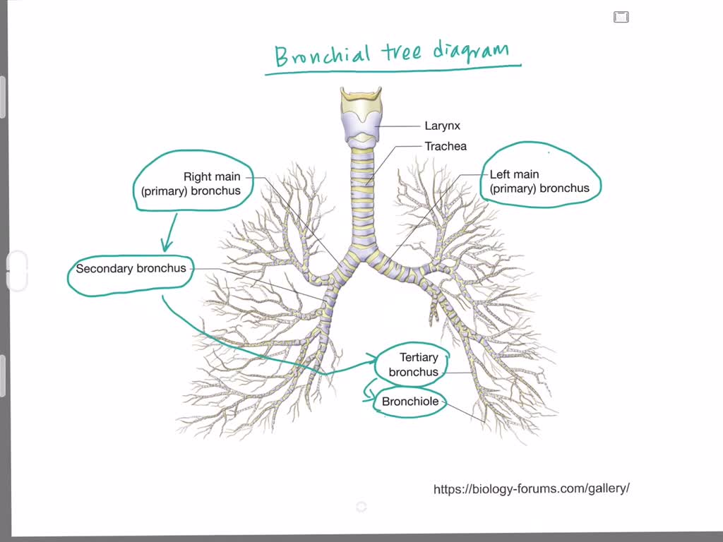

Macrophages, which engulf pathogens, are also present. Consists of respiratory bronchioles with alveoli attached to them.Įpithelial surfaces of airways up to respiratory bronchioles have cells that secrete mucus to trap particulate matter in the air, which is then moved by cilia present on these cells and swallowed.

This consists of the tracheal tube, which branches into two bronchi. The conducting zone where there is no gas exchange.Airways beyond the larynx are divided into 2 zones: A respiratory cycle consists of an inspiration (inhalation) movement of air from the external environment into alveoli, and expiration (exhalation) – the movement of air from alveoli to the external environment.ĭuring inspiration, air passes through nose/mouth, pharynx (throat), and larynx. Airways are tubes through which air flows between the external environment and alveoli. Transport of H+ Ions Between Tissues and LungsĮach lung is composed of air sacs called alveoli – the sites of gas exchange with the blood.


 0 kommentar(er)
